
Research topics
Cell Membrane-inspired Phospholipid Polymers for Developing Medical Devices
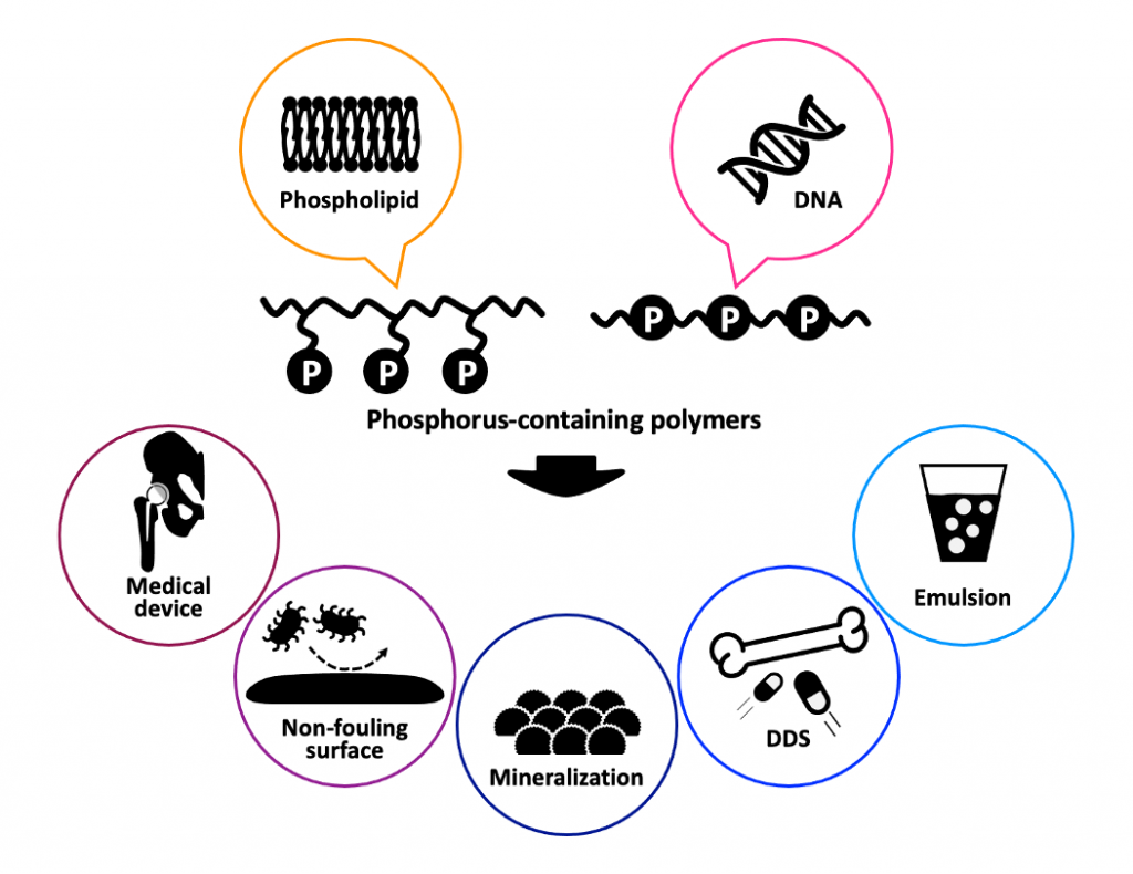
A new concept was proposed for designing blood-compatible polymeric materials with cell membrane-like surfaces; this concept uses 2-methacryloyloxyethyl phosphorylcholine (MPC), which is a methacrylate monomer with a phosphorylcholine (PC) group. MPC polymers can provide an artificial cell membrane structure at the surface and serve as excellent biointerfaces between artificial and biological systems. They have also been applied in the surface modification of some medical devices including long-term implantable artificial organs. An MPC polymer biointerface can suppress unfavorable biological reactions such as protein adsorption and cell adhesion – in other words, specific biomolecules immobilized on an MPC polymer surface retain their original functions. MPC polymers are also being increasingly used for creating biointerfaces with artificial cell membrane structures.
- Langmuir 2023;39:15417-15430.
- Colloids Surf. B 2021;250:111900.
- ACS Appl. Bio Mater. 2020;3:1071−1078.
Design of Drug Carriers with Biodegradable Polyphosphoesters for Targeting Therapeutics
Polyphosphoesters have been new candidates for polymeric biomaterials because of their biocompatibility and structural similarities to naturally occurring nucleic and teichoic acids. The phosphoester backbone is degradable through spontaneous hydrolysis and is accelerated with enzymatic treatment. Furthermore, a variety of polyphos- phoesters can be designed in comparison with conventional biodegradable polymers because cyclic phosphates as mono- mers are obtained from the condensation of alcohol and chloro-cyclic phosphate. We have recently performed the first instance of polyphosphoester synthesis using organocatalysts. According to this new process, high molecular weight polymers with good yield and narrow distribution could be obtained.
- Materialia 2024;36:102166.
- Adv. Funct. Mater. 2024:2316201.
- J. Biomater. Sci. Polym. Ed., 2023;34:2319-2331.
Cell Surface Modification
Methacryloyl groups are delivered on a living cell surface via a glycosylation pathway. The mannosamine derivative ManM is synthesized as a precursor of cell-surface sialic-acid residues. HeLa cells are cultivated in a culture medium containing ManMA, after which a sufficient amount of PEG410K-SH is in contact with the cells in the presence of a photoinitiator. The cells are then exposed to UV-light for 10 min. The immobilization of PEG410K-SH, termed PEGylation, on the cell surface is confirmed by fluorescence microscopy. The surface modification does not influence cell viability. Biotinylation of cell surface can also be achieved by the addition of a vinyl compound during PEGylation. By using this process, the functionalities of a cell surface can be freely controlled.